Lymphedema and lipedema
Two conditions that are often confused, yet they are not the same. Understanding the difference is the first step toward the right therapy.
What is lymphedema and what is lipedema
Lymphedema
Swelling caused by a disruption in the function of the lymphatic vessels. Positive Stemmer's sign, may involve the dorsum of the foot, carries a risk of erysipelas. Without therapy it progresses through stages.
Learn more →Lipedema (lipo-lymphedema)
Occurs mostly in women. Bilateral, symmetrical swelling from the iliac crest to the ankle joint. The dorsum of the foot is never involved.
- Stemmer's sign negative
- Soft skin texture, slight pitting or none on pressure
- No erysipelas
- Pain on palpation, bruises easily
Lymphedema — definition and classification
Lymphedema is a chronic, progressive condition of the body (arising from a disruption of the lymphatic system), which cannot be cured but can be fully kept under control with adequate treatment. Lymphedema is not an ordinary edema — with lymphedema, Stemmer's sign is positive, the foot is involved, and there is a risk of erysipelas.
Etiological classification (by cause)
Primary lymphedema: Arises from a congenital defect in the development of the lymphatic system, and can be congenital, early-onset or late-onset.
Secondary lymphedema: Develops later in life due to external factors such as surgery, radiation, injury or infection.
Clinical classification (by course)
Acute: Short-term swelling that resolves quickly.
Transient: Time-limited swelling that subsides.
Chronic: A long-lasting, persistent condition that requires treatment.
Stages of chronic lymphedema
Stage 0 (latent): The swelling is not yet visible, even though the lymphatic system is already damaged and functioning inadequately.
Stage 1 (spontaneously reversible): The swelling is soft, pressure leaves a pit, and elevating the limb reduces the swelling.
Stage 2 (spontaneously irreversible): The swelling is firmer, tissue begins to bind (fibrosis), and elevating the limb no longer helps much.
Stage 3 (elephantiasis): The swelling is extreme, the skin is thickened with characteristic wart-like growths.
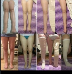
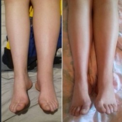
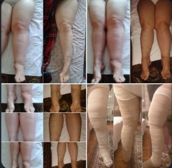
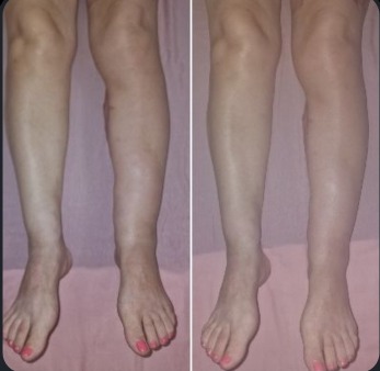
Leg lymphedema — clinical examples
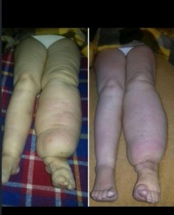
Leg lymphedema — progression through stages
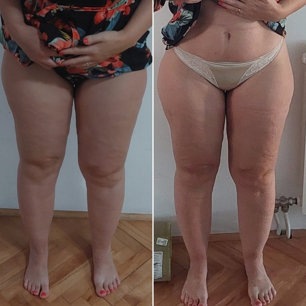
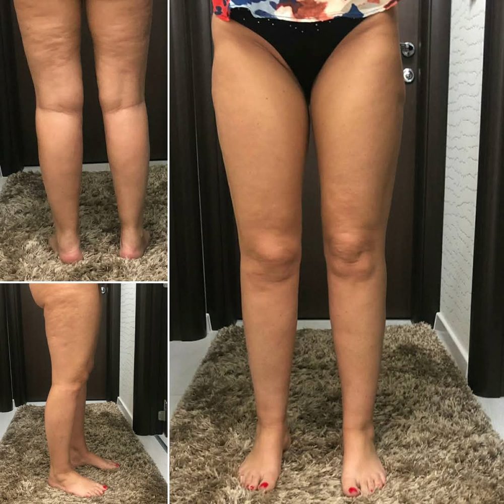
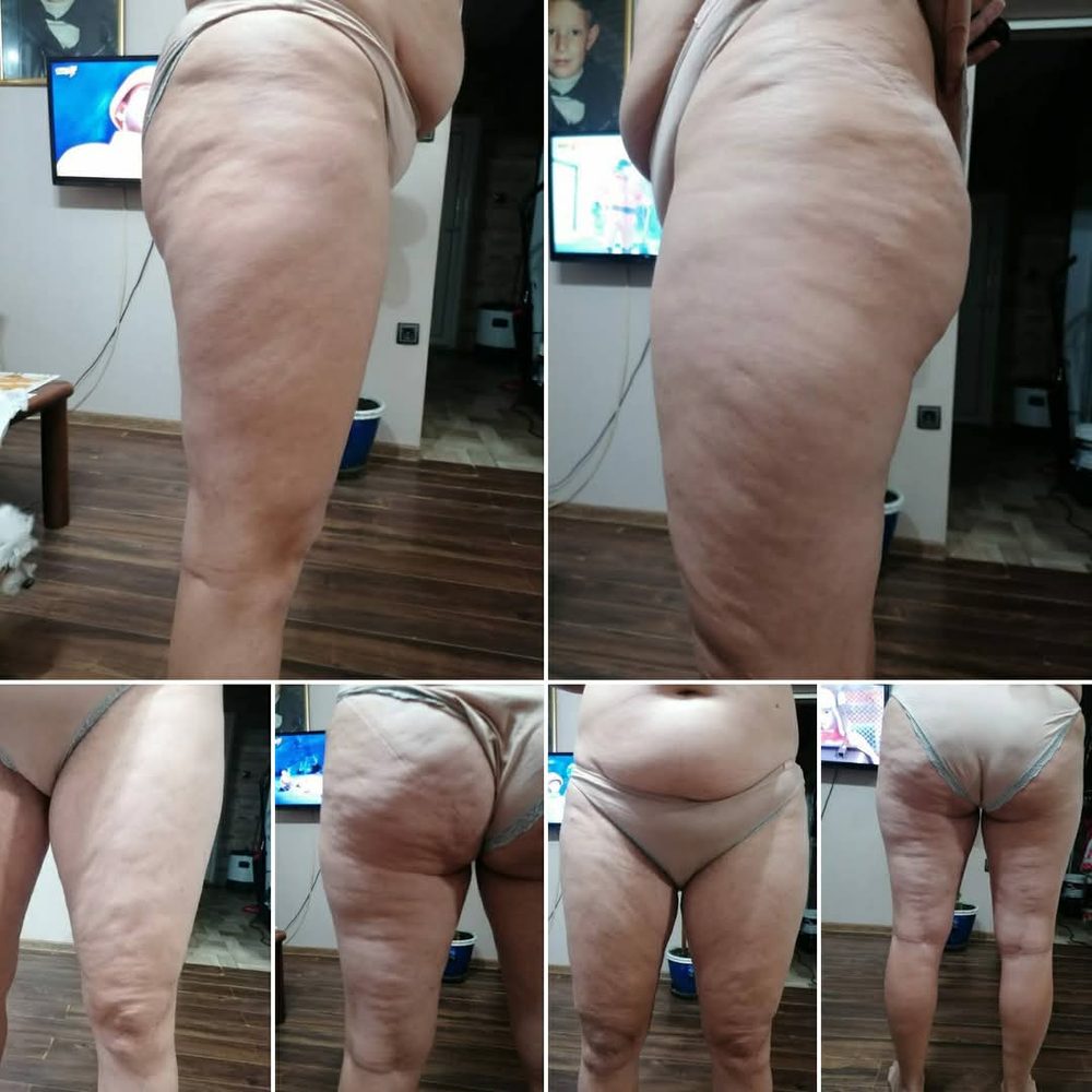
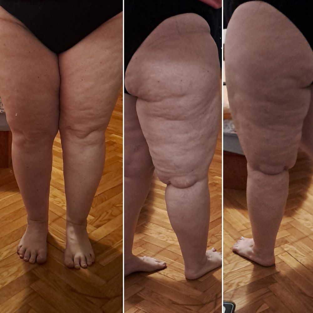
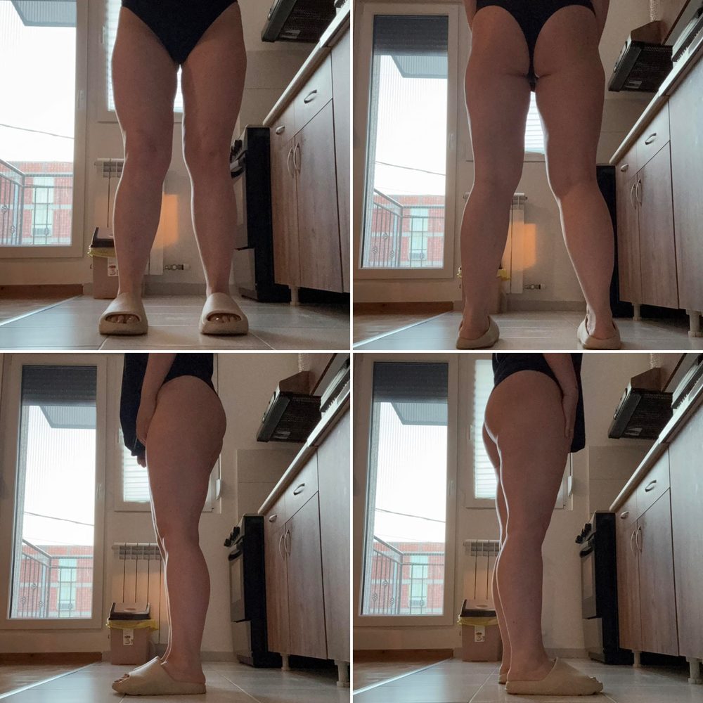
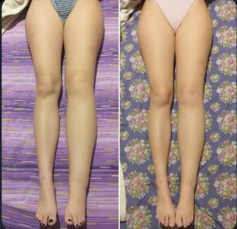
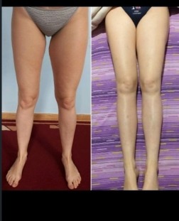
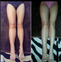
Leg lipedema — clinical examples
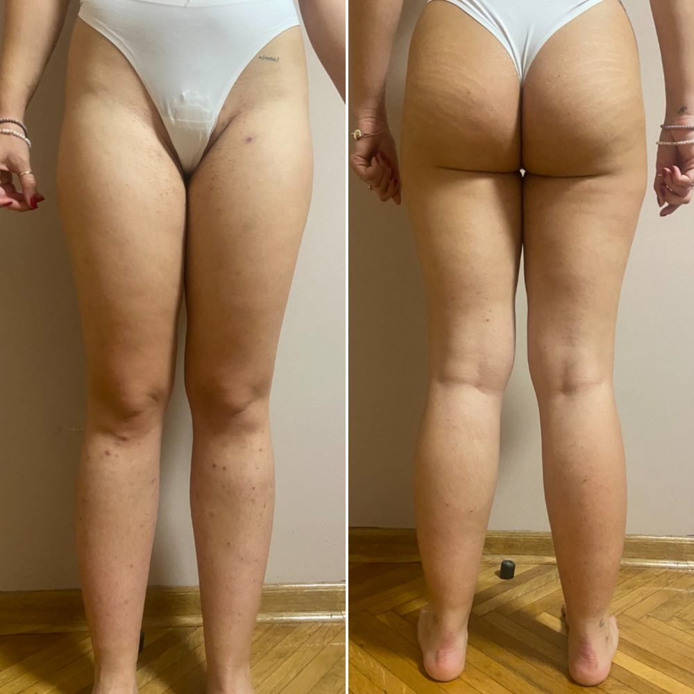
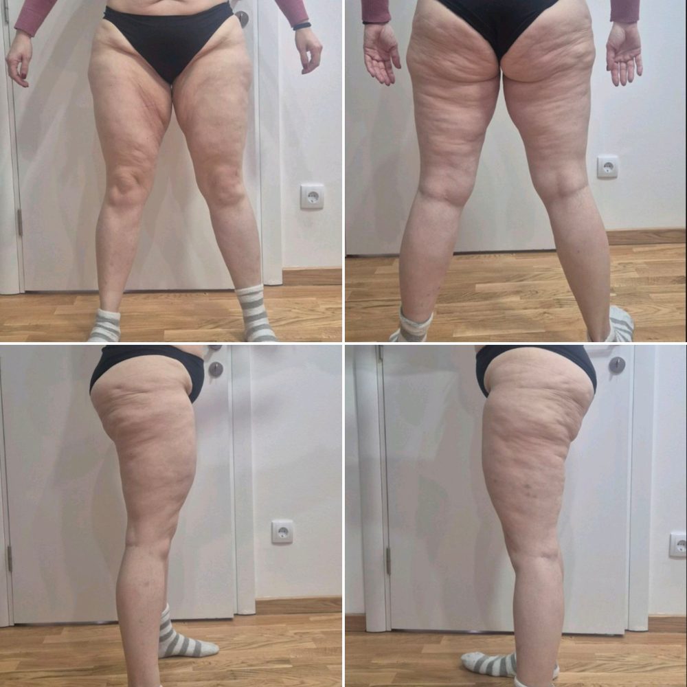
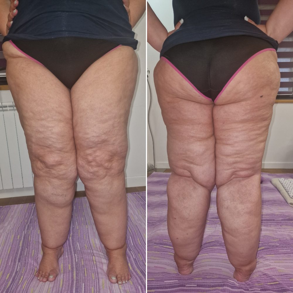
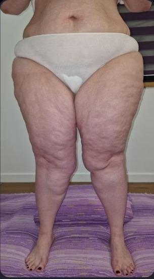
Leg lipedema — progression through stages
How to tell one from the other
| Characteristic | Lymphedema | Lipedema |
|---|---|---|
| Distribution | Most often unilateral | Bilateral, symmetrical |
| Dorsum of foot | May be involved | Never involved |
| Stemmer's sign | Positive | Negative |
| Skin on pressure | Pitting | Soft texture, slight or no pitting |
| Erysipelas | Risk exists | Not present |
| Pain | Usually painless | Pain on palpation |
| Bruising | Rare | Occurs easily |
| Sex | Both sexes | Almost exclusively women |
Lymphedema or lipedema? Check in one minute
Answer six questions about the swelling. Based on the differences in the table you will get a rough assessment.
Your answers are closer to the description of lymphedema.
Lymphedema is a chronic condition that cannot be cured, but with adequate treatment — manual lymphatic drainage, compression therapy and CDT — it can be kept fully under control.
Lymphedema therapy →Your answers are closer to the description of lipedema.
Lipedema (lipalgia syndrome) is painful fatty tissue that bruises easily and is resistant to diet and exercise. The modern approach is holistic: compression, manual lymphatic drainage, low-intensity movement and stabilizing body weight.
Lipedema treatment →Your answers show signs of both conditions.
Lipedema and lymphedema can also occur together (lipo-lymphedema). In such cases a professional examination is especially important.
Services overview →This is not a diagnosis. Only a professional examination can give an accurate assessment.
3 stages of lymphedema
Latent phase — the lymphatic system is already functioning inadequately, but no edema is present.
Learn more →Reversible phase — soft, protein-rich swelling, pressing with a thumb leaves a pit.
Learn more →Spontaneously irreversible phase — fibrosis and sclerosis, elevation no longer helps.
Learn more →Elephantiasis — extreme swelling, high risk of erysipelas.
Learn more →Primary lymphedema
Primary lymphedema is caused by a congenital malformation of the lymphatic vessels/nodes and in most cases occurs in the female population, where Stemmer's sign is positive in 90% of cases, as an indicator of already established fibrosis. From a pathological standpoint it occurs due to aplasia, hypoplasia or fibrosis of the lymph nodes. It is asymmetrical and localized in the peripheral parts of the limb. It can be spontaneous or hereditary in type.
Secondary lymphedema
Secondary lymphedema develops later in life due to external factors such as surgery, radiation, injury or infection.
Four stages of lipedema
The skin surface is smooth, the subcutaneous tissue is thickened and soft.
Learn more →The skin surface is uneven, firmer subcutaneous nodules, more pronounced swelling.
Learn more →Larger tissue deformations, pronounced folds and lumps.
Learn more →Lipo-lymphedema, a combination with lymphedema and additional swelling.
Learn more →Lipalgia syndrome (painful fat syndrome)
In professional circles, lipedema is increasingly referred to as lipalgia syndrome, because that name better describes the essence of the condition. It is not fluid-based swelling, but painful fatty tissue that bruises easily and is resistant to diet and exercise.
Liposuction is not a cure for this condition. It removes fatty tissue, but does not stop the underlying disorder. The modern approach is holistic: stabilizing body weight, compression therapy, manual lymphatic drainage, low-intensity movement and psychological support, while surgery is only considered as part of a broader plan.
Chronic venous insufficiency
Chronic venous insufficiency is a condition that can potentially cause lymphedema. The approach depends on the stage.
Prevention
Wearing a compression stocking is mandatory as the most important form of therapy. Manual lymphatic drainage can be performed preventively.
Learn more →Active therapy
Indication for manual lymphatic drainage and multi-layer bandaging (CDT).
Learn more →Erysipelas (cellulitis, red wind)
An acute streptococcal skin infection. In people with lymphedema it represents a serious complication and requires an urgent response. The condition is not contagious, and occurs sporadically, more often in colder periods.
The illness begins suddenly: a spike in temperature to 38–40°C (chills, fever, headache, malaise). Local signs appear after 12–24 hours: tightening of the skin, itching, then sharply demarcated redness that spreads quickly, along with swelling and skin that is warm, tense and painful to the touch.
Cause
Streptococcus
Duration (untreated)
15–25 days
Treatment
Causal (antibiotic) and symptomatic — under a physician's care
Note: this text is for informational purposes only and does not replace a medical examination.
See clinical example →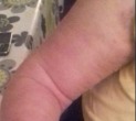
Erysipelas — clinical example
Characteristic sharply demarcated redness, swelling and skin that is tense and painful to the touch — the typical appearance of erysipelas (red wind) on the arm of a person prone to lymphedema.
General tips for preventing lymphedema
- Keep the limb elevated above heart level.
- Impeccable daily skin hygiene, with low-pH lotions free of alcohol, dyes, lanolin, petroleum derivatives and perfume, to prevent bacterial and fungal infection — pay special attention to folds and the areas between the toes/fingers.
- Avoid minor injuries: use an electric shaver for hair removal, wear gloves when working, use a thimble when sewing, take care of your nails and don't cut cuticles, wear closed shoes and cotton socks, keep feet always clean and dry.
- Avoid working with sharp objects — needles, scissors, awls, glass, thorns.
- Clean any scratch or burn with soap and water and treat it promptly.
- Watch for signs of infection — redness, warmth, swelling, pain — and see a doctor immediately.
- It is forbidden to give injections or vaccines, draw blood, perform lymphography or acupuncture, shave with a blade, remove hair in a way that irritates the skin, use rough massage technique, remain in a forced position for a long time, or fly on a plane without bandaging or compression garments on the affected limb.
- Tan gradually, in the shade, using creams with a high UV protection factor.
- Avoid extreme heat or cold — ice or hot packs, saunas, hot baths, tanning beds, direct heat sources, drying hair under a hood dryer; shower with lukewarm water and use air conditioning on hot days.
- See a doctor for any kind of skin rash.
- Do not overload the affected limb — carry out long-lasting work, pushing, pulling and carrying loads exclusively with the healthy arm, and no heavier than 2.5–3 kg.
- Avoid applying excessive external pressure: wear loose clothing and shoes, avoid jewelry and necklaces, don't sit with crossed legs, don't sleep on the affected limb, don't measure blood pressure on the affected arm, wear an appropriate orthopedic bra.
- Do not sit with your legs down in one position for longer than 30 minutes.
- Avoid standing still for a long time — it's better to alternately raise and lower the feet, shift body weight from leg to leg, from heel to toes.
- Walking, swimming, light aerobics and cycling are recommended.
- Regular check-ups with your doctor at scheduled appointments.
- Check the entire limb every day; measure its circumference and see a doctor if it worsens.
- The sense of touch, temperature or pain may be weaker in the affected region, so use the unaffected limb to check bath water temperature or for other purposes (e.g. checking milk before giving it to a child).
Ready to take the first step?
At the examination we assess your condition together and build a therapy plan tailored just for you.
Book an examination